Much of the work that has been accomplished on the assessment of serum progesterone in normal pregnancy was done from the early 1950’s through the 1970’s. Furthermore, most of the assessment of progesterone in pregnancy as it relates to various complications of pregnancy was accomplished from the early 1960s through the early 1980s. In spite of improvements in the accuracy and precision of progesterone assays since that time and a better ability to date pregnancy and establish more accurate gestational ages, very little subsequent work has been accomplished in this area.
However, data on the level of progesterone in normal pregnancy, and as it relates to a variety of pregnancy-related complications and features of previous reproductive history has been generated in a study which was conducted from the years 1980 through 2001 at the Pope Paul VI Institute. Modern means of progesterone assessment with improved accuracy and precision were used along with more precise means of dating the pregnancies.
In this study, 610 patients through 830 pregnancies and 8,545 progesterone levels were studied and statistically evaluated. These patients were primarily infertility patients who were receiving progesterone supplementation during the course of their pregnancy. Infertility was either primary or secondary and some of the patients also had a history of previous spontaneous abortion and, in some cases, recurrent spontaneous abortion.
The complete details of this study have been published in the textbook, The Medical & Surgical Practice of NaProTECHNOLOGY. The physician reader of this web site should consult that textbook for all of the details. A summary of the statistically significant changes in progesterone levels by condition or event is presented in Table 54-1.
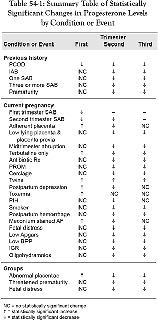
It has been known for a long time that progesterone is decreased during the first trimester of pregnancy in patients who have spontaneous abortions. It has also been thought that the placenta takes over the production of progesterone during the second and third trimesters of pregnancy. This study shows that the level of progesterone in first and second trimester spontaneous abortions, are indeed statistically decreased. In fact, this observation supports the structure of the study conducted at the Pope Paul VI Institute.
However, a decrease in progesterone production, presumably by the placenta, during the second and third trimester of pregnancy in a variety of different pregnancy-related complications and previous pregnancy historical events was also observed. This suggests that the role of progesterone as an indication of placental function may be more significant than what had been previously appreciated and is especially important into the second and third trimesters of pregnancy. This stage of pregnancy has only been minimally studied in the past. While it is not new that a variety of pregnancy-related complications are associated with decreased progesterone production, in the study conducted at the Pope Paul VI Institute, there was an expanded evaluation of that very type of assessment. For example, it has been shown previously that serum progesterone levels have been noted to be decreased in intrauterine death, premature labor, threatened premature labor, premature rupture of the membranes, amnionitis and abruption of the placenta. Increased levels of progesterone have also been observed in twin pregnancies, Rh isoimmunization and hydatidiform mole. The last two conditions were not a component of the study at the Pope Paul VI Institute.
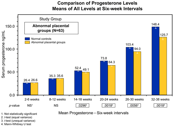
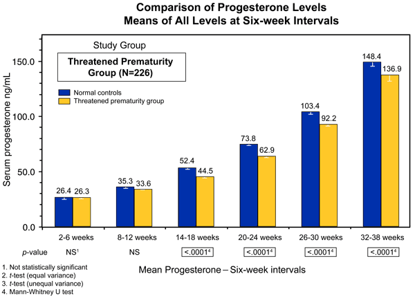
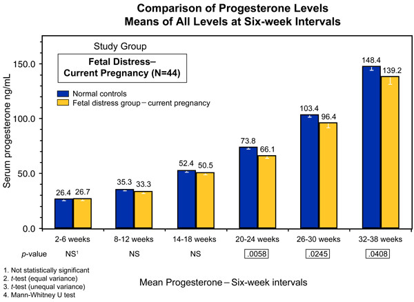
The data in the Institute’s very large and systematic assessment of progesterone in pregnancy suggests that the more significant progesterone-deficient time period in pregnancy is actually after the first trimester and into the second and third trimester. This is noted by comparing the sum of the pooled progesterone levels collected at six-week intervals during the course of pregnancies in a group of patients who had abnormal placentae, threatened prematurity and fetal distress (Figures 54-9, 54-10 and 54-11).
Progesterone Support During Pregnancy
Progesterone support in pregnancy has been in use for nearly 60 years, having received its start with publications dating back to the 1940s. Its initial use was in patients who had habitual spontaneous abortion caused by luteal phase deficiency. Luteal phase deficiency is due to a failure of the function of the corpus luteum in the production of progesterone from the corpus luteum is indispensable during the first seven weeks of pregnancy. Surgical removal of the corpus luteum during this period of time results in pregnancy loss and progesterone replacement can help maintain the pregnancy. There is evidence of support in the concept that progesterone given in early pregnancy may be useful in some women with recurrent miscarriage and that the measurement of serum progesterone levels in early pregnancy can be an adjunctive marker for the further assessment of pathologic pregnancies.
The administration of progesterone later in pregnancy has been considered to be justified because of an observed decrease in circulating progesterone with the onset of labor, and association of premature labor with decreased progesterone concentrations and the observation that progesterone has a tocolytic effect. It is thought that the administration of exogenous progesterone might, therefore, reduce uterine contractions and help prevent preterm labor.
This idea has received a considerable boost from the recent wide-spread publicity given to two papers which showed a significant reduction in preterm delivery rates with the prophylactic administration of either progesterone or 17 alpha-hydroxyprogesterone caproate. While this was portrayed as a “major breakthrough” by the national media, in reality, the use of progesterone (or 17 alpha-hydroxyprogesterone caproate) for the prevention of preterm labor has appeared in the medical literature for nearly 30 years.
Progesterone itself, rather than any of its metabolites, appears to be the active, natural progestational compound in the uterus. In conjunction with estradiol, progesterone has the following important functions:
- It stimulates the growth of the uterus
- It causes “maturation” (i.e., differentiation) of the endometrium, converting it to a secretory type.
- It stimulates the decidualization of the endometrium required for implantation
- And it inhibits myometrial contractions.
A thorough discussion of this topic is presented, in detail, in the textbook, The Medical & Surgical Practice of NaProTECHNOLOGY and the physician reader of this web site is specifically referred to that text for more information.
It might be of some interest to the reader to note that the retroplacental blood pool contains progesterone levels that are three to six times the maternal level during late pregnancy. In addition, fetal serum levels are seven times the maternal levels. Thus, the fetus is exposed to very high concentrations of progesterone in the natural state, much higher than could be accomplished even with the exogenous administration of progesterone.
The Pope Paul VI Institute now has over a 25-year experience in the use of progesterone support in pregnancy and the basic protocol is presented here, however, the details must be reviewed in the textbook, The Medical & Surgical Practice of NaProTECHNOLOGY, prior to implementation.Back to Top
Indications for the Use of Progesterone
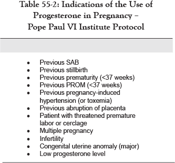
In Table 55-2, the various indications for the use of progesterone in pregnancy with the use of the Pope Paul VI Institute protocol, is listed.
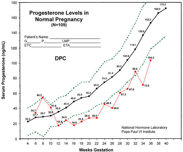
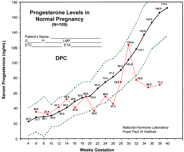
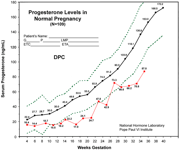
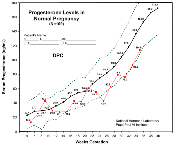
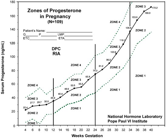
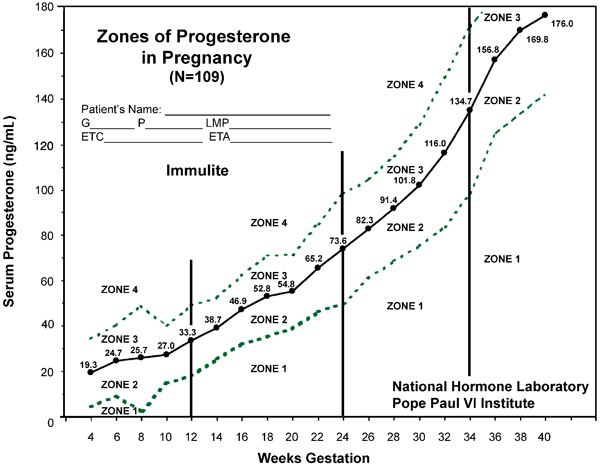
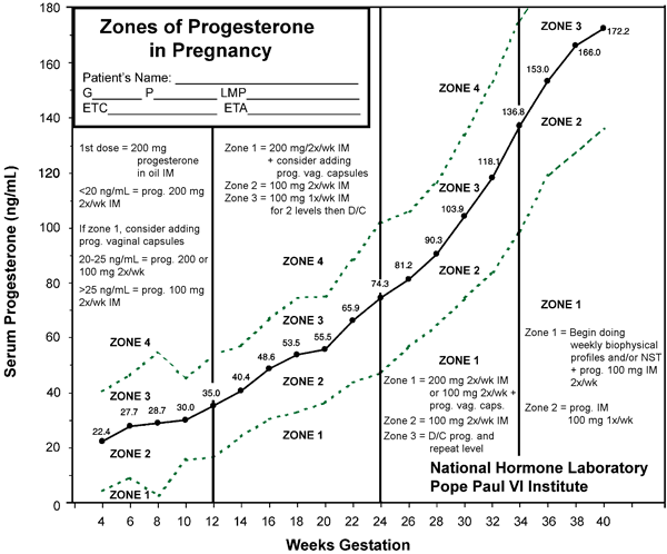
With the study of progesterone that has been ongoing at the Pope Paul VI Institute, a protocol for the utilization of progesterone has been developed. Serum progesterone levels are monitored on a two-week basis and applied to the normogram which is presented in Figure 55-4 and 55-5. These two curves using radioimmunoassay or the Immulite system of Diagnostic Products Corporation are virtually identical. The serum progesterone level is divided into four separate zones and the levels can be plotted during the course of the pregnancy.
The exact protocol for the implementation of progesterone and its use during the course of pregnancy, is outlined in a schematic fashion in Figure 55-6.
Safety of Progesterone in Pregnancy
There has been, over the years, a good deal of commentary that suggests progesterone is, in some way or another, associated with fetal abnormalities when used in pregnancy. This question is examined in detail in the textbook, The Medical & Surgical Practice of NaProTECHNOLOGY. This includes the 25-year experience of the Pope Paul VI Institute and a report of the outcomes of 933 pregnant patients who received progesterone during the course of their pregnancy. This is the largest single study of its kind ever conducted. The incidence of fetal abnormalities was actually lower in that population than it was in the population that did not receive progesterone.
The conclusion reached upon extensive study of this topic at the Pope Paul VI Institute, specifically as it relates to the naturally-occurring hormone progesterone, is the following: There is no credible evidence to suggest that its use to support pregnancy, whether that support be in the early days or months of pregnancy or later in pregnancy, is, in any way, teratogenic or responsible for any genital malformations. In fact, all of the available evidence strongly supports it safety when used in pregnancy.
In this report, over 2,000 pregnancies were reviewed or reported with the use of progesterone support in pregnancy with no increase in birth defects or genital anomalies. Progesterone support in pregnancy can be considered completely safe!
A few examples of the utilization of progesterone and its monitoring during the course of pregnancy are shown in Figures 55-9, 55-10, 55-11 and 55-12.
